
.jpg)

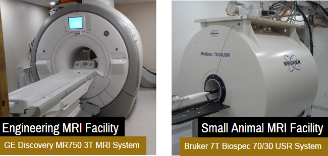


Practical Small Animal MRI is the seminal reference for clinicians using Magnetic Resonance Imaging in the diagnosis and treatment of veterinary patients. Although MRI is used most frequently in the diagnosis of neurologic disorders, it also has significant application to other body systems. Magnetic resonance imaging (MRI) is a very exciting and clinically rewarding diagnostic aid used at Animal Imaging since 2004. Magnets commonly used in human and veterinary medicine range from 0. Magnetic resonance imaging (MRI) At Donnington Grove we are equipped with an onsite Magnetic Resonance Imaging (MRI) scanner. This is a very modern diagnostic tool available in only a few practices in this country. Practical Small Animal MRI is the seminal reference for clinicians using Magnetic Resonance Imaging in the diagnosis and treatment of veterinary patients. Although MRI is used most frequently in the diagnosis of neurologic disorders, it also has significant application to other body systems. The Small Animal Imaging Facility is a Stanford School of Medicine service center, and is supported by the Stanford Cancer Institute, as well as user fees levied on each instrument. It is operated by the Departments of Pediatrics and Radiology. The Small Animal Imaging Facility (SAIF) is a core facility of the University of Pennsylvania and provides multimodality radiological imaging and image analysis for cells, tissues, and small animals. Magnetic resonance imaging (MRI) produces highcontrast, anatomically detailed tomographic images without the use of xrays. Although MRI has traditionally been used for diagnosing various diseases affecting the central nervous system, in recent years the range of clinical applications for MRI has broadened considerably. Hallmarq's PetVet MRI scanning is a powerful tool designed to provide easy diagnosis for companion animals. Take a look at our PetVet MRI videos here. A stateoftheart MRI machine, offering the clearest images yet to aid in treating everything from neurological conditions to cancer, will soon make its home at NC State. NC State Veterinary MRI replaces the MRI service AnimalScan presently used by the hospital. Magnetic Resonance Imaging (MRI) is a noninvasive imaging test used to diagnose, and help aid in the treatment of, a variety of medical conditions in the head, neck, brain, joints and spine. Small Animal MRI Veterinary MRI scanners for small animal applications provide imaging of the brain, spine and musculoskeletal systems. The Vet MR and Vet MR Grande provide a high quality solution for veterinary practices looking to perform MRI inhouse. Kopf MRI Stereotaxic Instruments are designed for use with specific animals. Our small animal MRI frames can be used with rat or mouse. Our large animal MRI frames are designed for use with cat, monkey and dog. Atlas of Small Animal CT MRI is a highly illustrated diagnostic imaging guide to common clinical disorders of dogs and cats. Contains over 3, 000 high quality CT, MRI and related diagnostic images Offers a unique approach emphasizing comparative imaging and pathologic correlation Handbook of Small Animal MRI will help you make the most of one of the greatest advancements in veterinary practice in recent years, resonance imaging. Those using the services of mobile scanners dedicated to veterinary use, as well as those in practices with their own MRI machine, will benefit from this book. The BioSpec from Bruker can perform successful small animal MRI. This multipurpose instrument is ideal for resonance imaging research. Animal surgery and related procedures for imaging studies, such as cranial surgery, tumor implantation, catheterization, or tail vein injection To learn more about our facility, click the items on the left, and see how we can help on your research. Atlas of Small Animal CT and MRI PDF. This book is intended for residents and specialists in most any clinical specialty, motivated veterinary students, and any practicing veterinarian who routinely refers patients for advanced imaging. Small animal imaging is increasingly recognized as an important facet of preclinical and translational cancer research. Perhaps most significant among the clear advantages of imaging experimental animals is that physiology, pathology and novel phenotypes can be understood in the most relevant milieu. These problems are also characteristic of clinical MRI, but are exacerbated in small animal MRM by the large gradients and small bores of the animal scanners. A further issue is the need to control biological motion, including that of the heart and lung, to avoid pronounced artifacts. Practical Small Animal MRI is the seminal reference for clinicians using Magnetic Resonance Imaging in the diagnosis and treatment of veterinary patients. Although MRI is used most frequently in the diagnosis of neurologic disorders, it also has significant application to other body systems. About Us: Advanced Animal Imaging provides stateoftheart imaging for animals through MRI, ECHO and Ultrasound. We strive to give you expert analysis, advanced technology, rapid report turn around time and the most affordable imaging within the region. Small animal resonance imaging (MRI) is not just used to inspect the brain and neurological system. It is a new type of hightech imaging in recent years. (MRI) can be used for almost any part of the animal body tomography. Practical Small Animal MRI is a timely addition to the everexpanding literature on veterinary imaging. Magnetic resonance imaging is considered the goldstandard in soft tissue imaging and this text is an excellent compilation of MRI images and anatomic illustrations. The MRI examination will be interpreted by a boardcertified veterinary radiologist. The written report will be sent to your referring veterinarian the next business day. Galen (Galen MRI Systems Limited) specializes in developing newgeneration MRI systems (dedicated breast MRI systems and dedicated small animal veterinary MRI systems) with machinelearning and A. capabilities that help medical professionals provide affordable and quality diagnisos to patients. Bruker BioSpin provides small animal MRI solutions for preclinical and molecular MR imaging research. By combining the latest rf coil and CryoProbe technology with high field superconducting or permanent desktop and unique software packages, our systems deliver high spatial resolution imaging of living organisms. Practical Small Animal MRI is the seminal reference for clinicians using Magnetic Resonance Imaging in the diagnosis and treatment of veterinary patients. Although MRI is used most frequently in the diagnosis of neurologic disorders, it also has significant application to other body systems. Our MRI core facility has strong capacity in human and nonhuman primate MRI and highresolution smallanimal MRI. Largebore Magnetic Resonance Imaging. The BIC houses two research dedicated largebore MRI scanners: a 3T Siemens Prisma and a 7T system (2018). Small animal MRI (SAMRI) technology now has become so refined that it can produce very detailed images of an animals brain or other organs. Small Animal Vital Signs Monitor MouseOx Plus Monitor MRI sensor with four clips and 5 of fiber optic cable and 15 of standard cable Custom versions with up to 7 of fiber optic cable and 30 of standard cable can be specialordered; please contact BIOPAC with specific requests. SMALL ANIMAL CLINICAL SCIENCES (fa) GENERAL INFORMATION: General anesthesia is required for all MRI examinations. All patients must arrive the day before the scheduled procedure. Galen (Galen MRI Systems Limited) specializes in developing newgeneration MRI systems (dedicated breast MRI systems and dedicated small animal veterinary MRI systems) with machinelearning and A. capabilities that help medical professionals provide affordable and quality diagnisos to patients. Atlas of Small Animal CT MRI is a highly illustratedguide to the common clinical disorders of dogs and cats that arenow routinely diagnosed using computed tomography and imaging. The Animal Imaging Shared Resource's Small Animal Imaging Service offers MRI, CT and PET services to CU Cancer Center member and nonmembers. Facility: Our facility is fully equipped for resonance imaging and proton spectroscopy (MRI 1HMRS) studies on small animals. Handbook of Small Animal Imaging: Preclinical Imaging, Therapy, and Applications (Imaging in Medical Diagnosis and Therapy) Apr 12, 2016 by George C. Ford Animal MRI Facility Mission of the Facility. The mission of the Biomedical Engineering Department in vivo high field Animal Magnetic Resonance Imaging (MRI) Facility is to provide intellectual support and state of the art MRI facilities to researchers in the biomedical sciences and engineering who wish to image or collect nuclear resonance (NMR) spectra from small animals or. The use of small animal models for the study of normal development, of progression of human disease, and of treatment of human disease, has become now more easier with. The Small Animal Imaging Facility at The University of Texas MD Anderson Cancer Center sports three MRI scanners in its instrument stable, and they are by far the most widely used of the lab. Different dedicated small animal coils and several imaging sequences were evaluated to optimize image quality with respect to SNR, contrast and spatial resolution. As an application, optimal greywhitematter contrast and resolution were investigated for rats. Furthermore, manganeseenhanced MRI was. Small animal resonance microscopy (MRM) has evolved significantly from testing the boundaries of imag ing physics to its expanding use today as a tool in nonin Model 900M Small Animal MRI Stereotaxic Instrument is designed to successfully stabilize small animals for stereotaxic surgery including repositioning of the head holder assembly within a MRI device without generating interference. Practical Small Animal MRI is the seminal reference for clinicians using Magnetic Resonance Imaging in the diagnosis and treatment of veterinary patients. Preclinical imaging is the visualization of living animals for research purposes, such as drug development. Imaging modalities have long been crucial to the researcher in observing changes, either at the organ, tissue, cell, or molecular level, in animals. In this protocol, we will focus on small animal MR imaging and MR spectroscopy (MRIMRS) to noninvasively acquire relaxation weighted 1 H images of mouse and to obtain 31 P spectra of mouse muscle. This work does not attempt to cover every aspect of small animal MRIMRS but rather introduces basic procedures of mouse MRIMRS experiments. mri that fits your patients and your practice The PetVet is the only High Field (1. 5 Telsa) MRI system designed specifically for veterinarians. Features and benefits Practical Small Animal MRI is the seminal reference for clinicians using Magnetic Resonance Imaging in the diagnosis and treatment of veterinary patients. Although MRI is used most frequently in the diagnosis of neurologic disorders, it also has significant application to other body systems. Procedure Information: Magnetic resonance imaging (MRI) is a very exciting and clinically rewarding diagnostic aid used at Animal Imaging since 2004. MRI has been used medically for over 20 years and is now becoming available to veterinarians in select areas. The Small Animal MRI at Baylor College of Medicine provides training, instrumentation, technical expertise, and software for small animal MRI and image processing. This site provides information about specific equipment, services, fees, training, methods, and resources. Meanwhile, the Weldon School of Biomedical Engineering supports the Small Animal MRI Facility, which has a Bruker 7T Biospec small animal MRI system. This system is designed for small animals such as rodents, but it also is suitable for smaller samples and materials. The smallanimal system built at the University of Tbingen with this approach was also successfully used for in vivo studies, including cardiac gated simultaneous PETMRI acquisitions. A similar method was used to integrate a prototype human dedicated brain scanner with a 3T MRI scanner (Siemens Medical Solutions) ( 48 )..